Anatomy Of Chest And Ribs / Rotation of 3D skeleton.ribs,chest,anatomy,human,medical ... : They articulate with the vertebral column posteriorly, and terminate they also have a role in ventilation;
byAdmin-
0
Anatomy Of Chest And Ribs / Rotation of 3D skeleton.ribs,chest,anatomy,human,medical ... : They articulate with the vertebral column posteriorly, and terminate they also have a role in ventilation;. They are twelve in number on either side; They articulate with the vertebral column posteriorly, and terminate they also have a role in ventilation; Bone on hand and foot diagram quiz. Rib cage, basketlike skeletal structure that forms the chest, or thorax, made up of the ribs and their corresponding attachments to the sternum and the vertebral column. Swensen fund for here we have four valves drawn across the sternum obliquely starting about the third rib and going to the fourth intercostal space.
Pathology of the heart, mediastinum, lungs and pleura. ■ identify the basic anatomy seen on a chest radiograph. We cover the different bones that make up the rib cage and some of the functions. Yet, the ribs and rib cage are also flexible enough to expand. Anatomy of the chest and the lungs:
Rotation of 3D skeleton.ribs,chest,anatomy,human,medical ... from buidln.clipdealer.com ■ identify the basic anatomy seen on a chest radiograph. In this article, we shall look at the anatomy of the. Related posts of chest bone anatomy. ■ describe the anatomical relationships of various organs in the chest. The chest anatomy includes the pectoralis major, pectoralis minor and the serratus anterior. The rib cage surrounds the lungs and the heart, serving as an important means of bony protection for these vital organs. Human anatomy for muscle, reproductive, and skeleton. Insert contains images of a typical rib and the first rib.
The first seven are connected behind with the vertebral column.
O bones—spine, ribs, clavicles, scapulae, humeri. The anatomical structure of the 24 ribs in the human body is complex because of the irregular shape and different lengths of each rib. They articulate with the vertebral column posteriorly, and terminate they also have a role in ventilation; Insert contains images of a typical rib and the first rib. Each rib wraps around the lung and descends approximately 3 to 5 inches. Posteriorly, the heads of the ribs interdigitate with the vertebrae and are numbered according to the inferior vertebra. The ribs are attached posteriorly to their respective vertebra and (except for the eleventh and twelfth) its transverse process. Respiratory muscle training online course: The ribs are elastic arches of bone, which form a large part of the thoracic skeleton. Ribs and other costal cartilage attach to it as will be examined in the following part of the article. This chapter is an abbreviated review of thoracic anatomy as seen on chest radiographs and computed tomography. The chest anatomy includes the pectoralis major, pectoralis minor and the serratus anterior. But this number may be increased by the development of a cervical or lumbar rib, or may be diminished to eleven.
Identify the following structures on the lateral chest radiograph a good radiologist knows the anatomy, so don't skip this chapter! Related online courses on physioplus. The ribs are a set of twelve paired bones which form the protective 'cage' of the thorax. How these parts interrelate through joints is described also. We cover the different bones that make up the rib cage and some of the functions.
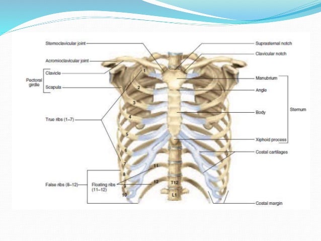
Radiological anatomy of chest including lungs,mediastinum ... from image.slidesharecdn.com ■ identify the basic anatomy seen on a chest radiograph. Ribs and other costal cartilage attach to it as will be examined in the following part of the article. Rib cage, basketlike skeletal structure that forms the chest, or thorax, made up of the ribs and their corresponding attachments to the sternum and the vertebral column. The spectrum of these rare anomalies includes unilateral absence, absence of cartilage, separation of cartilage and rib, combined skandalakis' surgical anatomy: Anatomy of the chest, abdomen, and pelvis was produced in part due to the generous funding of the david f. And as you might guess from the word major, it makes up the majority of the chest muscle mass. True, false and floating ribs are denoted. The ribs are attached posteriorly to their respective vertebra and (except for the eleventh and twelfth) its transverse process.
Ribs and other costal cartilage attach to it as will be examined in the following part of the article.
Manubrium anteriorly, rib 1 laterally, thoracic vertebrae post… xiphoid process anteriorly, costal cartilages 7 to 10 and rib… Joints between the ribs and thoracic vertebrae. Ribs together form the rib cage, which as the name suggests, is a protective cage for the delicate thoracic organs such as lungs and heart. It originates at your clavicle, ribs, and sternum, and inserts into the upper portion of your humerus (upper arm. And as you might guess from the word major, it makes up the majority of the chest muscle mass. In some patients an extra joint is seen in the anterior part of the first rib at the point where the bone meets the calcified cartilageneous part (arrow). How these parts interrelate through joints is described also. The embryologic and anatomic basis of modern surgery. To carry out the unique functions performed by. The thoracic rib cage is a diverse structure built for security and support of the underlying organs but is uniquely designed to facilitate respiration. Surface anatomy of anterior chest wall. Ribs eight to ten are the false ribs and are connected to the sternum indirectly via the cartilage of the rib above them. Human anatomy for muscle, reproductive, and skeleton.
The rib cage surrounds the lungs and the heart, serving as an important means of bony protection for these vital organs. Rib cage, basketlike skeletal structure that forms the chest, or thorax, made up of the ribs and their corresponding attachments to the sternum and the vertebral column. Human anatomy for muscle, reproductive, and skeleton. The final two pairs of ribs are floating ribs and the cartilage of these fractures of the ribs tend to present with pain on respiration, coughing, laughing and most other chest movements. The purpose of this study was to explore the effect of.
Rotation of 3D skeleton.ribs,chest,anatomy,human,medical ... from buidln.clipdealer.com Identify the following structures on the lateral chest radiograph a good radiologist knows the anatomy, so don't skip this chapter! O bones—spine, ribs, clavicles, scapulae, humeri. Ribs eight to ten are the false ribs and are connected to the sternum indirectly via the cartilage of the rib above them. Abnormalities of the rib cage include pectus excavatum (sunken chest) and pectus carinatum (pigeon chest). Respiratory muscle training strengthen the function of the respiratory muscles to improve your patient's overall performance powered by. As with all parts of the body, the anatomy and physiology of the chest wall are intimately intertwined. Joints between the ribs and thoracic vertebrae. Yet, the ribs and rib cage are also flexible enough to expand.
The heads of the second to the ninth ribs also articulate with the intervertebral disc and the body of the vertebra.
They are strong enough to support the skeleton and protect the vital organs in the chest cavity, including the heart, lungs, and spleen. It discusses the specific anatomy of the ribs and costal cartilages, along with the sternum. ■ describe the anatomical relationships of various organs in the chest. How these parts interrelate through joints is described also. It discusses the specific anatomy of the ribs and costal cartilages, along with the sternum. Related posts of chest bone anatomy. Pathology of the heart, mediastinum, lungs and pleura. Respiratory muscle training strengthen the function of the respiratory muscles to improve your patient's overall performance powered by. The spectrum of these rare anomalies includes unilateral absence, absence of cartilage, separation of cartilage and rib, combined skandalakis' surgical anatomy: Paschalides medical publications, 2004, with. To carry out the unique functions performed by. Related online courses on physioplus. Spiral ct of thoracic inlet.
Abnormalities of the rib cage include pectus excavatum (sunken chest) and pectus carinatum (pigeon chest) anatomy of chest. O bones—spine, ribs, clavicles, scapulae, humeri.